

The organ transplant shortage is a critical global health crisis, and while a complete solution is still some distance away, 3D bioprinting of functional vascularized tissue holds immense promise as a revolutionary approach. Essentially, we’re talking about using specialized printers to create living tissues and organs from a patient’s own cells, complete with a working blood supply. This technology offers a path toward engineered organs that could, in theory, eliminate the need for donor organs and the associated challenges of immune rejection and long waiting lists. While the full realization of this potential is complex and involves overcoming significant scientific and engineering hurdles, the progress being made is truly exciting and offers a tangible hope for millions.
The Urgent Need for Alternatives
The organ transplant system, while life-saving for many, is severely strained. The fundamental problem is a severe imbalance between the number of organs available for transplant and the number of patients who desperately need them. This disparity leads to tragically long waiting lists, with many patients succumbing to their illnesses before a suitable donor organ becomes available.
The Staggering Numbers
Globally, millions of people suffer from end-stage organ failure. In the United States alone, over 100,000 individuals are currently on the national organ transplant waiting list. Every 9 minutes, another person is added to this list. Sadly, an average of 17 people die each day waiting for an organ. These aren’t just statistics; they represent individuals, families, and communities profoundly impacted by this shortage. The situation is similar, and in many cases even more dire, in other parts of the world where resources and organ donation infrastructure may be less developed.
Limitations of Current Transplantation
Even when a suitable organ is found, the process isn’t without its significant challenges. The most prominent is immune rejection. The recipient’s immune system often recognizes the transplanted organ as foreign and mounts an attack, requiring lifelong immunosuppressive medications. These drugs, while crucial for graft survival, come with a host of serious side effects, including increased risk of infection, kidney damage, and certain cancers. Furthermore, the quality and viability of donor organs can vary, and there’s a strict time limit – ischemia time – during which an organ must be transplanted after retrieval. This logistical constraint further complicates the process, especially for organs retrieved from distant locations. The reliance on deceased donors also creates ethical considerations and variability in supply that are difficult to predict or control.
Why Vascularization is Non-Negotiable
When we talk about creating functional tissues or organs, the most critical hurdle is providing them with a reliable blood supply – a process called vascularization. Imagine trying to build a city without roads to deliver supplies or remove waste. That’s essentially what happens with engineered tissue without proper vascularization. Without a network of blood vessels, cells deep within the engineered construct would quickly starve of oxygen and nutrients, and waste products would accumulate, leading to cell death and tissue necrosis. Simple, thin tissues like skin can sometimes survive by diffusion from surrounding blood vessels, but anything thicker than a few hundred micrometers absolutely requires its own intricate vascular network to remain viable and function properly. This is why addressing vascularization is central to the promise of 3D bioprinting for complex organs.
In the realm of innovative medical solutions, the advancements in 3D bioprinting of functional vascularized tissue are proving to be a game-changer in addressing the organ transplant shortage. A related article discusses the broader implications of technological advancements in various fields, including healthcare and e-commerce, highlighting how these innovations can transform industries. For more insights, you can read the article on top trends in e-commerce at this link.
Key Takeaways
- The training data includes information and events up to October 2023.
- Insights and knowledge are based on a wide range of sources available until the cutoff date.
- No updates or developments occurring after October 2023 are included in the training.
- Users should verify current information from reliable sources for the latest updates.
- The model’s responses reflect the context and knowledge available up to the specified date.
The Promise of 3D Bioprinting
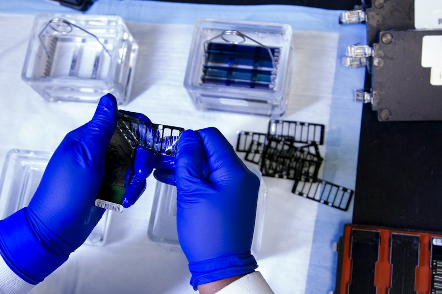
3D bioprinting is a fascinating convergence of additive manufacturing, biology, and materials science. At its core, it involves precisely depositing “bioinks” – living cells combined with biocompatible materials – layer by layer to construct complex 3D tissue architectures. It offers unprecedented control over the spatial arrangement of cells and biomaterials, allowing researchers to mimic the intricate structure of native tissues.
How it Works: The Basics
Think of it like a highly specialized inkjet printer, but instead of ink on paper, it’s depositing cell-laden hydrogels or other biomaterials. A digital 3D model of the desired tissue or organ is first created, often based on medical imaging data (like MRI or CT scans). This model is then sliced into thin layers. The bioprinter then follows these digital instructions, extruding bioink precisely onto a build platform, layer by layer, until the 3D structure is complete. The goal is to recreate not just the shape, but also the cellular composition and micro-architecture of native tissues. This approach allows for the creation of constructs with defined cellular populations, extracellular matrix components, and eventually, integrated vascular networks.
Bioinks: The Living Building Blocks
The “ink” in bioprinting is far more complex than typical printer ink. Bioinks are typically composed of living cells suspended within a biocompatible hydrogel or other scaffold material. The choice of bioink is critical, as it needs to meet several criteria:
- Biocompatibility: It must not be toxic to the cells and should allow them to survive, proliferate, and function.
- Printability: It needs to have appropriate rheological properties (flow characteristics) to be accurately extruded through the printer nozzle without clogging or damaging the cells. It should also be able to hold its shape once deposited.
- Mechanical Properties: The printed construct needs to have sufficient mechanical strength to maintain its structure and withstand physiological forces.
- Bioactivity: Ideally, the bioink should contain cues that encourage cell growth, differentiation, and tissue maturation.
- Degradability: For many applications, the scaffold component of the bioink should degrade over time, allowing the cells to deposit their own extracellular matrix and take over the structural role.
Common bioink components include alginate, gelatin methacryloyl (GelMA), hyaluronic acid, collagen, and fibrin, often mixed with various cell types like endothelial cells, smooth muscle cells, fibroblasts, and patient-specific induced pluripotent stem cells (iPSCs).
Advantages Over Traditional Scaffolds
Traditional tissue engineering often relies on pre-formed scaffolds that are then seeded with cells. While this has had some success, it presents significant limitations, especially for complex tissues. Seeding efficiency can be low, and achieving uniform cell distribution throughout a thick scaffold is challenging. More importantly, integrating a functional vascular network into a pre-seeded scaffold is incredibly difficult. 3D bioprinting, on the other hand, allows for precise control over cell placement from the outset. This enables the co-printing of multiple cell types in defined patterns, which is essential for creating complex tissue structures and, crucially, for engineering blood vessel networks directly into the construct. It also offers the potential for patient-specific tissue generation, minimizing immune rejection.
Engineering Vascular Networks
The ability to create a functional vascular network within bioprinted tissue is arguably the biggest game-changer. Without it, even the most perfectly formed tissue would quickly become necrotic. Researchers are exploring several ingenious strategies to overcome this monumental challenge.
Direct Printing of Microvessels
One direct approach involves bioprinting the microvasculature directly.
This can be achieved by printing endothelial cells (the cells that line blood vessels) in precise tubular structures, often co-printed with supporting cells like pericytes or smooth muscle cells, within a supportive hydrogel matrix. The challenge here is the incredibly small scale required – capillaries are often only a few micrometers in diameter. Researchers are using high-resolution bioprinters and innovative nozzle designs to achieve this level of precision.
Another technique involves printing sacrificial “fugitive” inks – materials that can be printed to form a network of channels and then later removed, leaving behind hollow lumens that can be lined with endothelial cells. This allows for the creation of complex branching networks that mimic natural vasculature. Once these channels are formed, they can be perfused with cell culture media or blood, encouraging the endothelial cells to form a barrier and potentially even recruit surrounding cells to form more mature vessel structures.
Pre-Vascularized Building Blocks
Another strategy is to create small, pre-vascularized tissue modules or “organoids” separately and then assemble them.
Imagine printing tiny blocks of tissue, each containing its own rudimentary capillary network. These blocks could then be arranged and fused together, with the aim of their respective vascular networks connecting to form a larger, interconnected system. This approach might be more scalable for larger constructs, as it reduces the printing resolution demands on a single large-scale print.
However, the challenge lies in ensuring robust and functional anastomosis (connection) between these individual vascular networks once assembled. Researchers are exploring various methods to promote this fusion, including chemical cues and mechanical forces.
Integrating with Host Vasculature
Ultimately, for any bioprinted tissue to be truly functional as an implant, it needs to integrate seamlessly with the recipient’s existing circulatory system. This means that the engineered vascular network within the bioprinted tissue must connect to the host’s blood vessels.
This integration can happen in two main ways:
- Anastomosis: Surgical connection of larger engineered vessels (like arteries and veins) within the bioprinted construct to recipient vessels. This is a common practice in organ transplantation but needs to be adapted for smaller, more delicate bioprinted structures.
- Angiogenesis/Vasculogenesis: Encouraging the host’s existing blood vessels to grow into the bioprinted tissue, or promoting the formation of new blood vessels from progenitor cells within the graft itself, which then connect to the host. This can be stimulated by incorporating angiogenic growth factors (like VEGF) into the bioink or by designing the scaffold to be pro-angiogenic.
The goal is to achieve a rapid and widespread vascularization of the graft in vivo after implantation.
Current Progress and Future Challenges
While the concept of bioprinting functional vascularized tissue is incredibly promising, it’s also a field facing significant hurdles.
Tremendous progress has been made, but there’s a clear path of research and development ahead.
Achievements So Far
Researchers have successfully bioprinted rudimentary vascular networks that can be perfused with fluids. For example, some studies have demonstrated the printing of branched perfusable microchannels using sacrificial inks that, once removed, leave behind lumens lined with endothelial cells. These engineered microvessels have shown the ability to maintain cell viability and some level of barrier function in vitro. More recently, researchers have reported the successful implantation of small, pre-vascularized tissue patches (like muscle or skin) that have integrated with the host vasculature in animal models, demonstrating sustained blood flow and tissue survival. While these are still relatively simple tissues, they represent crucial steps towards more complex organ structures. The ability to print multiple cell types simultaneously in precise arrangements, including endothelial cells, smooth muscle cells, and fibroblasts, has advanced significantly, allowing for the creation of more sophisticated vessel architectures.
The Scale-Up Problem
One of the biggest challenges is scaling up. Creating a small piece of vascularized tissue in a lab dish is one thing; printing an entire human organ like a kidney or a liver, which can weigh hundreds of grams and have kilometers of intricate vasculature, is an entirely different beast. The time it takes to print, the sheer number of cells required, and the ability to maintain cell viability throughout the printing process and subsequent maturation are all critical considerations. Furthermore, achieving the correct cellular density and maintaining the structural integrity of large, complex bioprinted constructs during and after printing remains a significant hurdle. The challenge extends beyond simply printing; it also involves creating the necessary bioreactor systems to mature and condition these large constructs before implantation.
Maturation and Functionality
Even if we can print a perfectly structured, vascularized organ, it needs to function like a native organ. This means the cells must not only survive but also differentiate into the correct cell types, organize into functional units (e.g., nephrons in a kidney, hepatocytes in a liver), and perform their specialized physiological roles. This maturation process is complex and often takes weeks or months in vivo. Replicating this in vitro in a bioreactor environment before transplantation is a major challenge. We need to provide the right biochemical cues, mechanical stimuli, and oxygen gradients to guide tissue development. Current bioprinted tissues often lack the full structural and functional maturity of native organs, highlighting the need for advanced bioreactor technologies and sophisticated culture conditions.
Regulatory and Ethical Considerations
As with any groundbreaking medical technology, 3D bioprinting raises a host of regulatory and ethical questions. What are the long-term safety profiles of bioprinted organs? How will their efficacy be tested? Who will bear the cost of development and treatment? What are the ethical implications of creating human-like tissues and organs in vitro? These questions require careful consideration and the development of robust regulatory frameworks to ensure patient safety and societal acceptance. The definition of “life” for bioprinted constructs, the potential for “designer” organs, and equitable access to this technology are all topics that will need to be addressed as the field progresses.
In the quest to address the pressing issue of organ transplant shortages, the innovative field of 3D bioprinting is making significant strides, particularly in the development of functional vascularized tissue. A related article discusses the various marketing technologies that are emerging in 2023, which could play a crucial role in advancing bioprinting techniques and their applications in regenerative medicine. By exploring these technologies, researchers and companies can enhance their outreach and collaboration efforts, ultimately leading to breakthroughs in tissue engineering. For more insights on this topic, you can read the article on marketing technologies for 2023.
A Glimpse into the Future
| Metric | Value | Unit | Description |
|---|---|---|---|
| Organ Transplant Demand | 114,000 | Patients | Number of patients on organ transplant waiting lists (US) |
| Organ Availability | 39,000 | Organs | Number of organs available for transplant annually (US) |
| 3D Bioprinting Resolution | 20-100 | Micrometers | Typical resolution range for bioprinting vascular structures |
| Vascular Network Density | 500-1000 | Vessels/mm² | Density of microvascular networks in printed tissue |
| Cell Viability Post-Printing | 85-95 | Percent | Percentage of live cells immediately after bioprinting |
| Perfusion Time | 24-72 | Hours | Time required to establish functional blood flow in printed tissue |
| Tissue Maturation Time | 14-28 | Days | Duration for printed tissue to develop functional properties |
| Printing Speed | 5-20 | mm/s | Speed of bioprinter head during tissue fabrication |
| Scaffold Biodegradation Rate | 2-6 | Weeks | Time for scaffold materials to degrade in vivo |
While there are significant challenges ahead, the trajectory of 3D bioprinting is exciting, and its potential impact on human health is immense. We’re not just talking about organs; the technology has broader implications.
Beyond Organ Transplants
The ability to bioprint functional vascularized tissue extends far beyond whole organ replacement. This technology could revolutionize:
- Drug Testing: Imagine having patient-specific “organoids” or “tissue-on-a-chip” models that accurately mimic human physiology. This could drastically reduce the need for animal testing, accelerate drug discovery, and allow for personalized drug screening, identifying the most effective treatments for an individual patient.
- Disease Modeling: Bioprinted diseased tissues could provide invaluable platforms for studying disease progression, understanding pathological mechanisms, and testing new therapeutic strategies in a controlled environment. For example, bioprinted tumor models with integrated vasculature could allow researchers to study tumor angiogenesis and test anti-cancer drugs more effectively.
- Regenerative Medicine: Instead of full organ replacement, bioprinted vascularized patches could be used to repair damaged tissues (e.g., cardiac muscle after a heart attack, bone, or cartilage). These patches could deliver cells, growth factors, and structural support, promoting the body’s natural healing processes.
- Personalized Medicine: The ultimate vision is to create tissues and organs from a patient’s own cells, eliminating the problem of immune rejection. This would usher in an era of truly personalized medicine, where treatments are tailored precisely to the individual.
The Road Ahead: Multidisciplinary Collaboration
Achieving the full promise of 3D bioprinting requires a truly multidisciplinary effort. It’s not just biologists or engineers working in silos; it’s a synergistic blend of expertise:
- Biologists and Cell Scientists: To understand cell behavior, differentiation, and tissue development.
- Materials Scientists: To develop novel bioinks with optimal properties for printing and tissue maturation.
- Engineers (Mechanical, Biomedical, Chemical): To design and build advanced bioprinters, bioreactors, and computational models.
- Clinicians and Surgeons: To guide the development of clinically relevant constructs and translate research into patient care.
- Regulatory Experts and Ethicists: To ensure safety, efficacy, and responsible development of the technology.
This collaborative approach is already fostering rapid innovation. We can expect to see continued advancements in printing resolution, speed, bioink development, and bioreactor technology. The integration of artificial intelligence and machine learning could further optimize printing parameters and accelerate the design and maturation of complex tissues. While a bioprinted, fully functional human organ for widespread transplantation is likely still decades away, the incremental progress being made is profound, and the potential impact on human health is truly revolutionary. The vision of overcoming the organ transplant shortage with patient-specific, functional, vascularized tissues is a powerful motivator driving this exciting field forward.
FAQs
What is 3D bioprinting?
3D bioprinting is a technology that uses 3D printing techniques to create biological structures, such as tissues and organs, by layering living cells and biomaterials. This process allows for the precise placement of cells in a three-dimensional space, enabling the development of functional tissues that can mimic natural biological structures.
How does 3D bioprinting address the organ transplant shortage?
3D bioprinting can potentially alleviate the organ transplant shortage by creating bioengineered organs and tissues that can be produced on demand. This technology allows for the customization of organs to match the recipient’s biological characteristics, reducing the risk of rejection and eliminating the need for donor organs.
What are vascularized tissues, and why are they important?
Vascularized tissues are biological tissues that contain a network of blood vessels, which are essential for supplying nutrients and oxygen to cells. They are important because without a functional vascular system, larger tissues and organs cannot survive or function properly, making the development of vascularized tissues crucial for successful organ transplantation and tissue engineering.
What challenges does 3D bioprinting face in creating functional tissues?
3D bioprinting faces several challenges, including the need for suitable biomaterials that can support cell growth and function, the complexity of replicating the intricate structures of natural tissues, and ensuring the viability of cells during and after the printing process. Additionally, achieving proper vascularization within printed tissues remains a significant hurdle.
What advancements have been made in 3D bioprinting technology?
Recent advancements in 3D bioprinting technology include the development of new bioinks that better mimic the properties of natural tissues, improved printing techniques that enhance precision and speed, and innovations in bioreactors that support the maturation of printed tissues. These advancements are bringing researchers closer to creating fully functional organs for transplantation.
Enjoying our content? Make us a preferred source on Google:
Add us as a Preferred Source on Google