

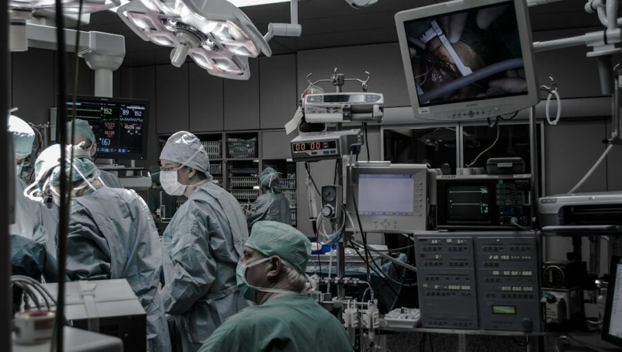
Ever wonder if surgeons could “practice” on a patient before the actual operation? Well, in a way, they can. Mixed Reality (MR) surgical pre-planning uses 3D holograms to let surgeons visualize and manipulate anatomical structures in a virtual space, significantly improving operative accuracy and patient outcomes. It’s not science fiction anymore; it’s a powerful tool changing how complex surgeries are approached.
Think of Mixed Reality as a blend of the real and virtual worlds. Unlike Virtual Reality (VR), which completely immerses you, or Augmented Reality (AR), which overlays digital information onto your real view, MR allows you to interact with virtual objects as if they were physically present in your real environment.
In surgical pre-planning, this means surgeons wear a headset that projects highly detailed 3D holographic models of a patient’s anatomy directly into their line of sight. These models are generated from standard medical imaging like CT scans and MRIs, transformed into interactive, volumetric representations.
The Problem with Traditional Pre-Planning
Historically, surgical planning relied heavily on 2D images. Surgeons would look at stacks of CT or MRI slices, mentally reconstruct the 3D anatomy, and then draw their incision lines or plan their approach. This method, while effective for many procedures, has its limitations.
- Mental Reconstruction is Challenging: Our brains are amazing, but visualizing complex 3D structures from 2D images is prone to error and can be difficult, especially for intricate anatomies or unusual pathologies.
- Lack of Spatial Awareness: It’s hard to accurately judge depth, distance, and relationships between structures when you’re just looking at flat pictures.
- Limited Collaboration: While surgeons can discuss cases over 2D images, the ability to collectively manipulate and interact with a 3D model together offers a whole new level of shared understanding.
How MR Transforms Pre-Planning
Mixed Reality bridges this gap by providing an intuitive, interactive 3D environment. Surgeons can walk around a holographic projection of a patient’s heart, liver, or brain, zoom in on specific areas, virtually dissect layers, and even “ghost” through tissues to see underlying structures.
- Enhanced Spatial Understanding: Seeing the anatomy in true 3D, to scale, in your own environment provides an unparalleled sense of spatial awareness. You can literally walk around the patient’s anatomy, examining it from every angle.
- Improved Anatomical Comprehension: Complex relationships between blood vessels, nerves, tumors, and organs become immediately clear. This clarity is crucial for avoiding critical structures during surgery.
- Interactive Manipulation: The ability to rotate, scale, segment, and even virtually “cut” into the holographic model allows for a dynamic planning process that simply isn’t possible with 2D images.
In the realm of advanced surgical techniques, the integration of mixed reality technologies is revolutionizing pre-operative planning and execution. A related article discusses the extended early bird pricing for a mobility conference, which highlights the importance of innovative technologies in healthcare. For more insights on how emerging technologies are shaping the future of surgery, you can read the article here: Mobility 2021 Early Bird Price Extended for One More Day.
Key Takeaways
- The training data includes information and events up to October 2023.
- Insights and knowledge are based on a wide range of sources available until the cutoff date.
- No updates or developments occurring after October 2023 are included in the training.
- Users should verify current information from reliable sources for the latest updates.
- The model’s responses reflect the context and knowledge available up to the specified date.
The Journey from Scan to Hologram
Getting a patient’s internal anatomy into a holographic format isn’t magic, but it does involve some clever technology. It’s a multi-step process that ensures accuracy and fidelity.
Image Acquisition
It all starts with high-quality medical imaging. CT (Computed Tomography) and MRI (Magnetic Resonance Imaging) scans are the primary sources. These imaging modalities capture a series of 2D slices of the body, which collectively contain the 3D information.
- CT Scans: Excellent for visualizing bone, dense tissues, and contrast-enhanced blood vessels. They provide fine spatial resolution.
- MRI Scans: Superior for soft tissue differentiation, making them ideal for brain, spinal cord, and joint imaging, as well as tumor detection.
Segmentation and 3D Reconstruction
Once the scans are acquired, the raw image data needs to be processed. This is where the heavy lifting happens in terms of converting 2D slices into a usable 3D model.
- Segmentation: This is the process of identifying and isolating specific anatomical structures (e.g., bones, organs, tumors, blood vessels) from the surrounding tissues within the 2D image slices. This can be done manually by skilled technicians, semi-automatically with user input, or increasingly, automatically using artificial intelligence (AI) and machine learning algorithms. Accurate segmentation is paramount, as any errors here will propagate into the 3D model.
- Surface and Volume Rendering: After segmentation, the isolated structures are reconstructed into 3D models. Surface rendering creates a geometric mesh representing the outer surface of an organ, while volume rendering displays the entire volumetric data, often with different colors or opacities to represent different tissue types. This step essentially “stacks” the segmented 2D slices to form a continuous 3D object.
Holographic Projection and Interaction
The final 3D model is then loaded into a Mixed Reality application. When a surgeon wears an MR headset (like Microsoft HoloLens), the device uses its cameras and sensors to map the real environment. The 3D model is then projected as a hologram into this real space, appearing as if it’s floating in front of the surgeon.
- Spatial Anchoring: The holograms are “anchored” to a specific point in the real world, meaning they stay in place as the surgeon moves around them. This is crucial for maintaining a stable and consistent view.
- Gesture and Voice Control: Surgeons interact with the holograms using natural gestures (e.g., pinching to zoom, swiping to rotate) or voice commands. This hands-free interaction is vital in a surgical context, where sterility and precise movements are key.
Key Benefits for Operative Accuracy

The primary advantage of MR surgical pre-planning is its profound impact on operative accuracy. This translates directly to better patient safety and outcomes.
Precision in Incision Planning
Deciding where to make the first cut is critical. With MR, surgeons can precisely plan the optimal incision location and length, considering underlying anatomy and minimizing unnecessary tissue damage.
- Optimal Entry Points: For minimally invasive procedures, finding the exact entry point for instruments is crucial.
MR allows surgeons to visualize the entire trajectory before making any incisions.
- Avoiding Critical Structures: By seeing nerves, blood vessels, and vital organs in 3D, surgeons can plan approaches that carefully navigate around these sensitive areas, reducing the risk of iatrogenic injury.
Enhanced Tumor Resection Strategies
Removing tumors completely while preserving healthy tissue is a delicate balance. MR provides unprecedented clarity for this task.
- Clear Margins: Surgeons can visualize the tumor in relation to surrounding healthy tissue, aiding in planning resection margins to ensure complete removal while minimizing damage to functional tissue.
- Complex Anatomy Navigation: For tumors nestled deep within complex organs (e.g., liver, brain), MR helps surgeons understand their precise relationship to major blood vessels and critical structures, informing the safest and most effective removal strategy.
Improved Trajectory Planning for Implants and Screws
In orthopedic and neurosurgical procedures, precise placement of implants or screws is paramount for stability and long-term success.
- Pre-visualization of Screw Paths: Surgeons can virtually “insert” screws into bone holograms, ensuring optimal length, angle, and avoiding vital structures like nerves or spinal cord.
- Custom Implant Fit: For custom-designed implants, MR can be used to visualize how the implant will fit within the patient’s unique anatomy, allowing for pre-operative adjustments if needed.
Expanding Applications and Future Horizons

While surgical pre-planning is a major use case, Mixed Reality’s potential in healthcare extends much further. The technology is still evolving, promising even more sophisticated capabilities.
Intraoperative Guidance (AR in the OR)
Beyond pre-planning, the next logical step is to bring these holographic models directly into the operating room as Augmented Reality (AR) overlays.
- “X-Ray Vision” During Surgery: Imagine a surgeon wearing an AR headset that superimposes the patient’s internal organs, tumors, and blood vessels directly onto their physical body during the actual operation. This provides “x-ray vision” for real-time guidance.
- Real-time Tracking: Advanced AR systems can track surgical instruments and the patient’s anatomy, dynamically updating the holographic overlay to maintain perfect registration even with subtle patient movements. This is still a developing field, but has immense potential.
Collaborative Planning and Training
MR isn’t just for individual surgeons. Its collaborative capabilities are transforming how medical teams work together and how new surgeons are trained.
- Multi-User Planning Sessions: Multiple surgeons, residents, and even referring physicians can simultaneously view and interact with the same holographic model, discussing surgical approaches and challenges in a shared virtual space. This fosters a deeper understanding and better-coordinated planning.
- High-Fidelity Surgical Training: Medical students and residents can practice complex procedures on realistic holographic anatomical models without any risk to patients. They can repeat procedures, explore different approaches, and receive real-time feedback in a safe, immersive environment.
Patient Education and Consent
Explaining complex surgical procedures to patients can be challenging using just diagrams or verbal descriptions. MR offers a powerful new tool for patient engagement.
- Visualizing the Procedure: Patients can see their own anatomy in 3D and watch a holographic simulation of the proposed surgery. This can significantly improve their understanding of the procedure, potential risks, and expected outcomes.
- Informed Consent: A better understanding of the surgery can lead to more truly informed consent, empowering patients to make confident decisions about their healthcare.
In the realm of advanced surgical techniques, the integration of mixed reality has shown remarkable potential in enhancing operative accuracy, as discussed in the article on Mixed Reality Surgical Pre-Planning: How 3D Holograms Improve Operative Accuracy. This innovative approach allows surgeons to visualize complex anatomical structures in three dimensions, leading to better pre-operative planning and execution. For those interested in exploring how technology can transform various fields, a related article on the best software for house plans provides insights into how 3D modeling is revolutionizing architectural design. You can read more about it here.
The Road Ahead: Challenges and Considerations
| Metric | Traditional Planning | Mixed Reality Surgical Pre-Planning | Improvement |
|---|---|---|---|
| Operative Accuracy (%) | 85 | 95 | +10% |
| Pre-Operative Planning Time (hours) | 4 | 2.5 | -37.5% |
| Intraoperative Time (minutes) | 120 | 90 | -25% |
| Surgeon Confidence Level (1-10) | 7 | 9 | +2 points |
| Postoperative Complication Rate (%) | 15 | 7 | -8% |
| Patient Recovery Time (days) | 14 | 10 | -29% |
While the benefits of Mixed Reality in surgery are clear, it’s not without its challenges. Adoption requires careful consideration of several factors.
Cost and Accessibility
MR headsets and the associated software and infrastructure can be expensive. Making this technology widely accessible to all healthcare institutions remains a hurdle.
- Hardware Costs: High-end MR headsets are still a significant investment for hospitals.
- Software Development and Integration: Creating robust, medically certified MR applications and integrating them with existing hospital IT systems requires specialized expertise and resources.
Workflow Integration
Integrating MR into existing surgical workflows requires careful planning and training. It’s not just about having the technology; it’s about seamlessly incorporating it into daily practice without disrupting established routines.
- Learning Curve: Surgeons and support staff need training to effectively use the MR hardware and software.
- Data Management: Securely managing and transferring sensitive patient data from imaging systems to MR platforms is crucial.
Regulatory Approvals and Clinical Validation
As a relatively new medical technology, MR surgical tools require rigorous testing, clinical validation, and regulatory approval to ensure their safety and efficacy.
- Clinical Trials: Demonstrating the tangible benefits of MR in terms of improved outcomes and reduced complications through well-designed clinical trials is essential for widespread adoption.
- Regulatory Oversight: Medical devices, especially those with such a direct impact on patient care, are subject to strict regulatory scrutiny by bodies like the FDA in the US or EMA in Europe.
Data Accuracy and Fidelity
The accuracy of the holographic models is entirely dependent on the quality of the initial medical images and the subsequent segmentation and reconstruction processes. Any errors or artifacts in these steps can lead to misleading holographic representations.
- High-Resolution Imaging: The better the quality and resolution of the CT/MRI scans, the more detailed and accurate the 3D models will be.
- Advanced Segmentation Algorithms: Continuous development of AI-powered segmentation tools is vital to improve accuracy and reduce the need for manual intervention, which can be time-consuming and operator-dependent.
Despite these challenges, the trajectory of Mixed Reality in surgery is undoubtedly upward. The clear advantages it offers in terms of improved operative accuracy, enhanced understanding, and better training are too significant to ignore. As the technology matures, costs decrease, and integration becomes more seamless, we can expect to see MR becoming an increasingly standard tool in the modern operating room, ultimately leading to safer and more effective surgeries for patients worldwide.
It’s an exciting time to be at the intersection of medicine and technology, and Mixed Reality is truly pushing the boundaries of what’s possible.
FAQs
What is mixed reality surgical pre-planning?
Mixed reality surgical pre-planning is a process that involves using 3D holograms and virtual reality technology to visualize a patient’s anatomy before a surgical procedure. This allows surgeons to plan and practice the operation in a virtual environment, improving accuracy and efficiency.
How do 3D holograms improve operative accuracy?
3D holograms provide surgeons with a detailed and interactive view of the patient’s anatomy, allowing them to identify potential challenges and plan the best approach for the surgery. By visualizing the anatomy in 3D, surgeons can make more precise decisions during the operation, leading to improved accuracy and outcomes.
What are the benefits of using mixed reality in surgical pre-planning?
Some benefits of using mixed reality in surgical pre-planning include enhanced visualization of the patient’s anatomy, improved communication among surgical teams, increased accuracy in planning and performing surgeries, reduced operating time, and better patient outcomes. It also allows for more personalized and tailored surgical approaches.
Is mixed reality surgical pre-planning widely used in the medical field?
While mixed reality surgical pre-planning is still relatively new, it is gaining popularity in the medical field. Many hospitals and surgical centers are starting to adopt this technology to improve surgical outcomes and enhance patient care. As the technology continues to advance, it is expected to become more widely used in the future.
Are there any limitations or challenges associated with mixed reality surgical pre-planning?
Some limitations and challenges of mixed reality surgical pre-planning include the initial cost of implementing the technology, the need for specialized training for surgical teams, potential technical issues during surgeries, and the time required to create and review 3D models. However, as the technology evolves and becomes more accessible, these challenges are expected to be addressed.
Enjoying our content? Make us a preferred source on Google:
Add us as a Preferred Source on Google