

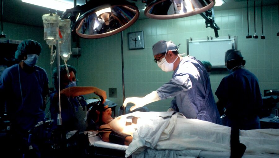
So, you’re curious about what AI is actually doing in the operating room, specifically when it comes to guiding surgeons? It’s not about robots wielding scalpels (yet!), but more about giving surgeons super-powered vision. This article dives into how AI, combined with fancy light technology called optical spectroscopy, is helping surgeons see and understand tissue in real-time during operations. Think of it as giving the surgeon an invisible, super-detailed x-ray vision, but instead of radiation, it’s using light. This helps them make better decisions, faster, which is pretty crucial when you’re, well, operating.
At its heart, AI-assisted surgical navigation with optical spectroscopy is about getting more information about the tissue a surgeon is working with, right when they need it. Traditional methods often rely on visual cues – what the tissue looks like, its texture, and how it feels. While experienced surgeons are amazing at this, there are limits to what the naked eye can discern. Optical spectroscopy changes that by shining specific wavelengths of light onto the tissue and then analyzing how that light bounces back or is absorbed. Different types of tissue, whether healthy or diseased, will interact with light in unique ways. AI then takes this complex light data and translates it into something meaningful for the surgeon.
What is Optical Spectroscopy, Anyway?
Imagine shining different colored lights on something and seeing how it reacts. Optical spectroscopy does something similar, but with a much wider range of light, including wavelengths we can’t even see, like infrared. When light hits tissue, some of it gets absorbed, some gets scattered, and some might even get re-emitted. The way this happens is heavily influenced by the chemical makeup and physical structure of the tissue.
How Does AI Come Into Play?
Raw spectroscopic data is a lot of numbers, often represented as complex graphs. For a surgeon in the middle of an operation, that’s not immediately helpful. This is where AI, particularly machine learning algorithms, shines. These algorithms are trained on massive datasets of spectroscopic data from known tissue types. They learn to identify the subtle patterns and signatures that distinguish, say, a healthy blood vessel from a tumor margin, or a nerve from surrounding connective tissue. The AI then processes the real-time spectroscopic readings from the patient’s tissue and instantly tells the surgeon, “This is likely healthy tissue,” or “This area shows characteristics of abnormal growth.”
AI-Assisted Surgical Navigation has been significantly enhanced by advancements in real-time tissue characterization using optical spectroscopy, which allows for more precise surgical interventions. For those interested in exploring the intersection of technology and everyday tools, a related article discusses the best tablets to buy for everyday use, which can be beneficial for medical professionals seeking portable devices for their practice. You can read more about it here: What is the Best Tablet to Buy for Everyday Use?.
Key Takeaways
- The training data includes information and events up to October 2023.
- Insights and knowledge are based on a wide range of sources available until the cutoff date.
- No updates or developments occurring after October 2023 are included in the training.
- Users should verify current information for accuracy beyond the training period.
- The model’s responses reflect the context and knowledge available up to the specified date.
Shining a Light on Tissue: The Spectroscopy Side
Optical spectroscopy isn’t a new concept; it’s been around for a while in labs and for analyzing materials. What’s new and exciting is how it’s being adapted and miniaturized for use directly within the surgical field. The goal is to provide immediate, objective information that complements the surgeon’s existing skills.
Different Types of Spectroscopy in Surgery
There are several ways spectroscopy can be used. Each has its own strengths and the choice often depends on the specific application and the type of information needed.
Near-Infrared Spectroscopy (NIRS)
NIRS is particularly useful because near-infrared light can penetrate tissues a few millimeters to a centimeter or so, allowing for the analysis of deeper structures without being overly invasive. It’s often used to monitor blood oxygen levels and blood flow, which are critical indicators of tissue health. Changes in these parameters can signal problems during surgery, like reduced blood supply to an area.
Raman Spectroscopy
Raman spectroscopy provides a detailed chemical fingerprint of the tissue. It’s highly sensitive to the molecular vibrations within the tissue, essentially revealing its biochemical composition. This can be incredibly powerful for distinguishing between different cell types or identifying specific molecules associated with diseases like cancer. Think of it as a super-specific chemical scanner.
Fluorescence Spectroscopy
This technique involves exciting the tissue with a specific wavelength of light and observing the light that the tissue emits in response (fluorescence). Different molecules in the tissue fluoresce differently. For instance, certain cancerous cells might have a different fluorescent signature than healthy cells, or surgeons might use fluorescent dyes that accumulate in specific tissues, and the spectroscopy then helps to precisely visualize those areas.
The Technology Itself: Probes and Systems
To get this light data from the patient, specialized probes are used. These are often small, fiber-optic based devices that can be attached to surgical instruments or held by the surgeon. The probe delivers the excitation light and collects the scattered or emitted light, sending it to a spectrometer. The spectrometer then analyzes the light and the processed information is displayed to the surgeon.
AI as the Interpreter: Making Sense of the Light Show

The real magic happens when the raw data from the spectrometer is fed into an AI system. Without AI, the data would be too complex and time-consuming for a surgeon to interpret in real-time. The AI acts as an incredibly sophisticated interpreter, translating subtle spectroscopic signatures into actionable insights.
Training the AI: Learning from Experience (Digitally)
The development of these AI systems involves extensive training.
Researchers and clinicians gather vast amounts of spectroscopic data from samples of known tissue types – healthy tissue, various stages of tumors, inflamed tissue, and so on. This data is meticulously labeled. The AI algorithms then learn to associate specific spectroscopic patterns with these known tissue types.
Real-Time Analysis: The Speed of Surgery
Once trained, the AI can analyze the spectroscopic data coming from the patient’s tissue in milliseconds.
This real-time capability is crucial. It means the surgeon receives immediate feedback as they are operating, allowing them to adjust their approach on the fly.
If the AI detects an area that is likely cancerous, the surgeon can ensure they have removed all of it.
If it indicates a critical nerve, they can be extra careful to avoid it.
Beyond Simple Identification: Predictive Capabilities
The AI isn’t just about saying “this is X” or “this is Y.” As the AI gets more sophisticated, it can start to offer predictive capabilities. For example, it might be able to predict how likely a particular margin is to have microscopic disease left behind, guiding the surgeon towards more aggressive removal if necessary.
Practical Applications in the Operating Room

The theoretical benefits of AI-assisted surgical navigation using optical spectroscopy translate into very tangible improvements in patient care. The primary goal is to enhance precision, reduce complications, and improve outcomes.
Cancer Surgery: The Frontier
One of the most promising areas is cancer surgery. Precisely identifying the full extent of a tumor and ensuring its complete removal (achieving clear margins) is critical for preventing recurrence.
Identifying Tumor Margins
During surgery, it can be visually challenging to distinguish the exact boundary between cancerous tissue and healthy tissue, especially for diffuse or infiltrating tumors. Optical spectroscopy, interpreted by AI, can highlight these subtle differences, helping surgeons to confidently excise all the tumor while sparing as much healthy tissue as possible. This can lead to better functional outcomes and reduce the need for repeat surgeries.
Sentinel Lymph Node Biopsy
In some cancer surgeries, surgeons need to identify and remove sentinel lymph nodes – the first lymph nodes that cancer cells are likely to spread to. Spectroscopy can potentially help identify these nodes more easily, reducing the invasiveness of the procedure and improving diagnostic accuracy.
Vascular and Reconstructive Surgery
Maintaining adequate blood flow is paramount in many surgical procedures. Spectroscopy can provide real-time monitoring of tissue perfusion.
Assessing Blood Flow and Oxygenation
In reconstructive surgery, such as skin grafts or free flap procedures, ensuring the blood supply to the transplanted tissue is vital for its survival. NIRS, for example, can monitor oxygen saturation in the graft in real-time. If oxygen levels drop, it signals a potential problem with the blood supply, allowing the surgeon to intervene before the tissue is irreversibly damaged.
Identifying Critical Vessels
During complex dissections, it can be difficult to differentiate between arteries, veins, and other critical vascular structures. Spectroscopy could potentially help in this differentiation, reducing the risk of accidental damage.
Minimally Invasive Surgery
As surgeries become more minimally invasive, surgeons are working with smaller incisions and often relying on laparoscopic cameras. This can limit their tactile feedback and visual cues. AI-assisted spectroscopy can bring enhanced information directly to these procedures.
Enhanced Visualization for Laparoscopy
A spectroscopic probe integrated into a laparoscopic instrument could provide real-time tissue characterization on the monitor, giving the surgeon an additional layer of information that is not readily available through standard video feeds.
AI-Assisted Surgical Navigation has seen significant advancements, particularly with the integration of real-time tissue characterization using optical spectroscopy. This innovative approach enhances the precision of surgical procedures by allowing surgeons to differentiate between healthy and diseased tissues during operations. For those interested in exploring related technological advancements in various fields, you might find this article on shared hosting services intriguing, as it highlights how cutting-edge technologies are transforming industries beyond healthcare.
Challenges and the Road Ahead
| Metric | Description | Value / Range | Unit | Notes |
|---|---|---|---|---|
| Spectral Resolution | Ability to distinguish between different wavelengths of light | 1-5 | nm | Depends on the optical spectrometer used |
| Real-Time Processing Latency | Time delay between data acquisition and output of tissue characterization | 50-200 | milliseconds | Lower latency improves surgical navigation accuracy |
| Tissue Classification Accuracy | Percentage of correctly identified tissue types | 85-95 | % | Varies with algorithm and tissue complexity |
| Penetration Depth | Depth at which optical spectroscopy can characterize tissue | 1-5 | mm | Depends on wavelength and tissue type |
| Number of Tissue Types Differentiated | Distinct tissue categories identified by the system | 5-10 | types | Includes healthy, malignant, and other tissue types |
| Signal-to-Noise Ratio (SNR) | Quality of the optical signal relative to background noise | 30-50 | dB | Higher SNR improves classification reliability |
| Integration with Surgical Navigation System | Compatibility and data synchronization capability | Yes | Boolean | Enables real-time guidance during surgery |
| Machine Learning Model Type | Algorithm used for tissue characterization | Random Forest / CNN / SVM | Type | Varies by implementation |
While the potential is immense, bringing AI-assisted optical spectroscopy into widespread surgical practice isn’t without its hurdles. It’s a complex technology that requires careful integration into the fast-paced and demanding environment of an operating room.
Standardization and Validation
One of the biggest challenges is ensuring that the AI models are robust and reliable across different patient populations, surgical settings, and even variations in equipment. Extensive clinical trials and validation studies are needed to prove efficacy and safety. Standardizing the way data is collected and processed is also crucial.
Integration into Workflow
Surgical workflows are highly optimized. New technologies need to be seamlessly integrated without adding significant time or complexity. This involves intuitive user interfaces and reliable hardware. The AI should augment, not disrupt, the surgeon’s established practice.
Cost and Accessibility
Advanced medical technologies often come with a significant price tag. Making these systems affordable and accessible to a wider range of hospitals and healthcare systems will be important for their broader adoption.
Regulatory Approval
Like all medical devices and software, AI-assisted surgical navigation systems will need to undergo rigorous review and approval by regulatory bodies such as the FDA. This process can be lengthy and demanding.
The Human Factor: Trust and Training
Ultimately, the surgeon is in charge. Building trust in the AI’s recommendations is key. This requires comprehensive training and a clear understanding of the system’s capabilities and limitations. The AI should be viewed as a powerful tool, not a replacement for the surgeon’s judgment.
Future Developments: Beyond Simple Identification
Looking ahead, the capabilities are likely to expand. We might see AI systems that can predict the likelihood of disease recurrence based on tissue characteristics, or even guide precise drug delivery to specific tissue types. The synergy between AI and optical spectroscopy is just beginning to unlock its full potential in revolutionizing how we approach surgery.
FAQs
What is AI-assisted surgical navigation?
AI-assisted surgical navigation refers to the use of artificial intelligence technology to help guide surgeons during procedures by providing real-time information and assistance.
How does optical spectroscopy contribute to tissue characterization during surgery?
Optical spectroscopy is a technique that uses light to analyze the chemical composition of tissues. By utilizing optical spectroscopy, surgeons can quickly and accurately characterize tissues in real-time during surgery.
What role does AI play in real-time tissue characterization during surgery?
AI algorithms are used to analyze the data collected through optical spectroscopy in real-time, providing surgeons with immediate feedback on tissue characteristics such as cancerous or healthy tissue, helping them make more informed decisions during the procedure.
What are the benefits of AI-assisted surgical navigation with optical spectroscopy?
Some benefits of AI-assisted surgical navigation with optical spectroscopy include improved accuracy in tissue characterization, reduced risk of errors during surgery, and potentially shorter operation times.
Are there any limitations or challenges associated with AI-assisted surgical navigation using optical spectroscopy?
Some limitations and challenges include the need for further validation of the technology, potential integration issues with existing surgical systems, and the requirement for specialized training for surgeons to effectively utilize the technology.
Enjoying our content? Make us a preferred source on Google:
Add us as a Preferred Source on Google